

 (03) 9455 1714


- About
- Laser vision correction
- Dry Eye
- Cross-Linking
About
Introduction

The cornea is responsible for two-thirds of the focusing power of the eye
The cornea is the transparent front part of the eye that covers the iris and pupil. The cornea is surrounded by the white of the eye (slera) and performs three vital functions.
- It provides a strong outer coat for the eye that protects it from trauma and infection.
- It is transparent allowing visible light but not dangerous UV light to pass through to the back of the eye.
- It maintains a very precise shape that refracts (bends) light so that a clearly focused image is seen on the retina. The cornea accounts for two-thirds of the eye’s total optical power. The lens, situated behind the iris, accounts for the remaining one-third.

The cornea needs to remain moist and will become painful, opaque and prone to infection if it dries out. Every time we blink the eyelids distibute tears across the cornea. Tears form in three layers, each of which must be healthy for optimum function.
- An outer, oily layer keeps tears from evaporating too quickly.
- A middle water layer makes up about 90% of the tear film and keeps the eye moist.
- A inner mucin layer helps fluid to remain on the surface of the cornea keeping it wet.
What can happen to the cornea?
The cornea can be affected by a number of conditions that can impact on you in terms of pain or loss of vision.
- Trauma can cause abrasions or damge to the surface of the eye causing pain, scarring or alteration in the shape of the cornea.
- Alterations in the shape of the cornea can cause refractive error and blurred vision.
- Infection can cause pain and subsequent scarring can cause opacification and loss of vision.
- A number of diseases can cause the cornea to lose transparency and blur vision.
- Exposure to UV light can cause growths from the edge of the cornea towards the centre of the eye, known as a pterygium.
Laser Vision Correction
Refractive Error
People may need glasses if the shape of their cornea does not accurately focus light onto the retina.
If the shape of your cornea is too steep light will focus in front of the retina (short-sighted). If the cornea is too flat, then light will focus behind the retina (long-sighted).

Eye without refractive error
Most people who have a refractive error need to wear glasses or contact lenses to see a clear image.

Laser vision correction
A high-tech laser is used to re-shape the cornea. The laser is extremely precise and is able to shape the cornea to within several thousandths of a millimeter.
Modern lasers can not only alter the shape of the cornea to correct long or short-sightedness but can also correct astigmatism at the same time.
LASIK Surgery
LASIK is the most common laser refractive procedure performed. A small flap is cut with a preliminary laser called the femtosecond laser. The flap is lifted allowing access to the underlying cornea. The extremely precise excimer laser is then able to re-shape the cornea without affecting the surface of the eye.

LASIK Surgery involves creating a flap
Is Laser Vision Correction Right For me
Who is suitable?
To be a suitable candidate for refractive surgery, you must be over 21 years of age. It is important that you have had no major change in your spectacle or contact lens prescription for at least 1 year.
Does it hurt?
LASIK is a popular choice for laser vision correction because visual recovery is rapid with minimal discomfort. Most patients can return to their normal activities very quickly.
Is it safe?
Laser vision correction is very safe with an extremely low rate of major complications. Dr Cumming will undertake an extensive and complete examination of your eyes and advise you of any risk involved in laser surgery as well as your chance of achieving clear vision without glasses.
Pterygium
What is Pterygium
Pterygium is an abnormal growth of the conjunctiva (skin covering the white of the eye) over the cornea (Clear part of the front of the eye). Pterygium can be unsightly and painful.
Symptoms of Pterygium
Mostly pterygium does not cause pain. However, it can appear unsightly if it is large and grows over more than a few millimeters of the cornea. A pterygium can become inflamed, if this happens it may become red and sore.
A pterygium that grows very large can alter the shape of the cornea and cause astigmatism. This can reduce your vision and it may be difficult to correct the severe astigmatism from a pterygium with glasses.

Pterygium can be unsightly and painful.
Non-Surgical options
Not all pterygiums require surgery. If the appearance does not bother you and it is not painful it may be best to leave it alone.
To minimise the risk of further growth and of the pterygium becoming inflamed it is important to use regular lubricants. Sun exposure should be avoided.
If the pterygium becomes red and painful you can use non-steroidal anti-inflammatory tablets or drops to help reduce the inflammation.

The pterygium is removed and graft site identified
Surgical options
Surgery is the only way to remove a pterygium and should be considered if:
- 1. The appearance bothers you
- 2. It is always or often painful
- 3. It is growing more than 2 mm onto the cornea
- 4. It is causing significant astigmatism
- 5. Prior to cataract surgery if it is large

Graft of healthy conjunctiva is sutured over the site of the pterygium
Pterygium Surgery
Surgery can be performed in a day theatre, a hospital or here in our treatment room at MCES.
Surgery involves removing the pterygium and taking a small patch of conjunctiva from under the upper eyelid. The conjunctival graft is then stitched or glued over the site of the pterygium to minimize the chance of re-growth.
What to expect after surgery
It is important to be aware that:
- your eye will be very sore for 1 week
- discomfort may persist for 1 month
The risks associated with surgery are minimal and your doctor will discuss these in detail. The greatest risk is that the pterygium will grow back, this can occur in 20% of cases.
Dry Eye
A thin fluid film protects the surface of the eye (cornea) from becoming dry.
The tear film has three important layers:
- A mucous layer at the bottom allows it to ‘gripʼ to small attachments on the cornea.
- A water layer or tear film forms the majority of the thin film.
- An oil layer protects the water from evaporation and helps the tear layer spread across the eye.

Tear film deficiency
The tear film is generally produced in very small amounts by cells that live on the white of the eye and under the eyelids.
With age these cells become less effective. Exposure to sunlight, dust, excessive heat and recurrent eye infections can further limit the ability of these cells to do their job. Drying of the tear film can cause two major problems.

Tired irritated eyes
If the cornea dries out it will start to rub against the underside of the eyelids and will develop lots of small abrasions. These can cause a sensation of tiredness, irritation, grittiness, or even pain. If left untreated this can lead to the possibility of a serious corneal infection.
Watery dry eye
Some people produce tears from the lacrimal gland in response to dry eye—the same as crying. The production of tears from the lacrimal gland is uncontrolled and results in a watery eye. The regular use of lubricating drops, at least four times a day, is the best treatment.
Oil layer deficiency
Some patients with dry eye have a healthy production of the tear film, but the overlying oil layer is weak or absent. This causes the tear layer to rapidly evaporate and inhibits it from spreading over the surface of the eye. Glands in the eyelids produce oil. These glands can become blocked in blepharitis.
Treatment
The regular use of a lubricant will help, but treatment should also be directed to improving the flow of oil from the glands in the eyelid.
Ask about IPL to treat blepharitis.
Herpetic Eye Disease
What is Herpetic Eye Disease
Herpetic eye disease is caused by a virus called Herpes Simplex Virus (HSV), the same virus that causes cold sores. In the eyes, it can cause infection or inflammation of the cornea.
Why does herpetic eye disease develop?
Many people have HSV living within their body. The virus lives in the nerve fibres of the body that supply the lips and eyes and generally donʼt cause any problems. Occasionally, the virus reactivates, and travels down the nerve fibres to start multiplying in the cornea or lips, causing herpetic eye disease or cold sores. This may happen when the bodyʼs immune system is weakened by some other health problem or stress.

Eye with Herpetic Eye Disease
How does herpes simplex virus affect the eye?
The cornea is most commonly affected by HSV. Infection affects the most superficial layer or skin of the cornea and is called epithelial keratitis. The bodyʼs immune system tries to kill the infection but sometimes this results in inflammation to the deeper layers of the cornea and is called stromal keratitis. Inflammation of these deeper layers is more serious because it can cause scarring of the cornea that can permanently blur your vision.

Detail of cornea showing patches of inflammation
Rarely HSV can cause inflammation of the iris and retina. Your eye may be dilated so that Dr. Joanne Goh can carefully check that your iris and retina are not affected.
Keratoconus and Cross-linking
Keratoconus
What is Keratoconus
The cornea is usually an even round shape. Keratoconus occurs when the cornea thins and bulges forward in a cone shape.
What causes keratoconus
We do not know the exact cause of keratoconus, but it is thought to have a genetic component and occurs more commonly in people with allergies like asthma or eczema. There is also an association between eye rubbing and keratoconus.

Normal vs Keratoconic Cornea
How keratoconus affects your vision
The cornea is important for focusing light onto the back of your eye. In keratoconus your vision becomes blurred because the cornea cannot focus light properly.

Blurring of image

Scar on cornea
Scarring of the cornea
Keratoconus may also cause scarring of the cornea, which can severely decrease your vision by blocking light from entering the eye
What is the natural history of keratoconus
Keratoconus is often diagnosed in young people in their teens or twenties. Keratoconus is a progressive condition and can gradually get worse with time. For many it stops progressing in their mid to late thirties.
Non-surgical treatment
In the early stages of the disease glasses can focus the light for you. In more advanced disease special contact lenses are required and in severe cases even this may not be enough to focus a clear image.
Corneal cross-linking
Corneal cross-linking is a treatment that can stop keratoconus from getting worse by strengthening the cornea. It can be performed in the clinic here at MCES and is successful in over 90% of patients. It can also be performed in hospital if you have private Health insurance, at Manningham Private Hospital.
Surgical treatment
If it is no longer possible to give you clear vision with glasses or contact lenses, then you may require a surgical procedure to regain clear vision:
- Laser eye surgery
- Intra-corneal implants
At your consultation with Dr Kathy Jao will carry out a comprehensive assessment and will determine the procedure that is most likely to give you the best outcome.
Cross-linking
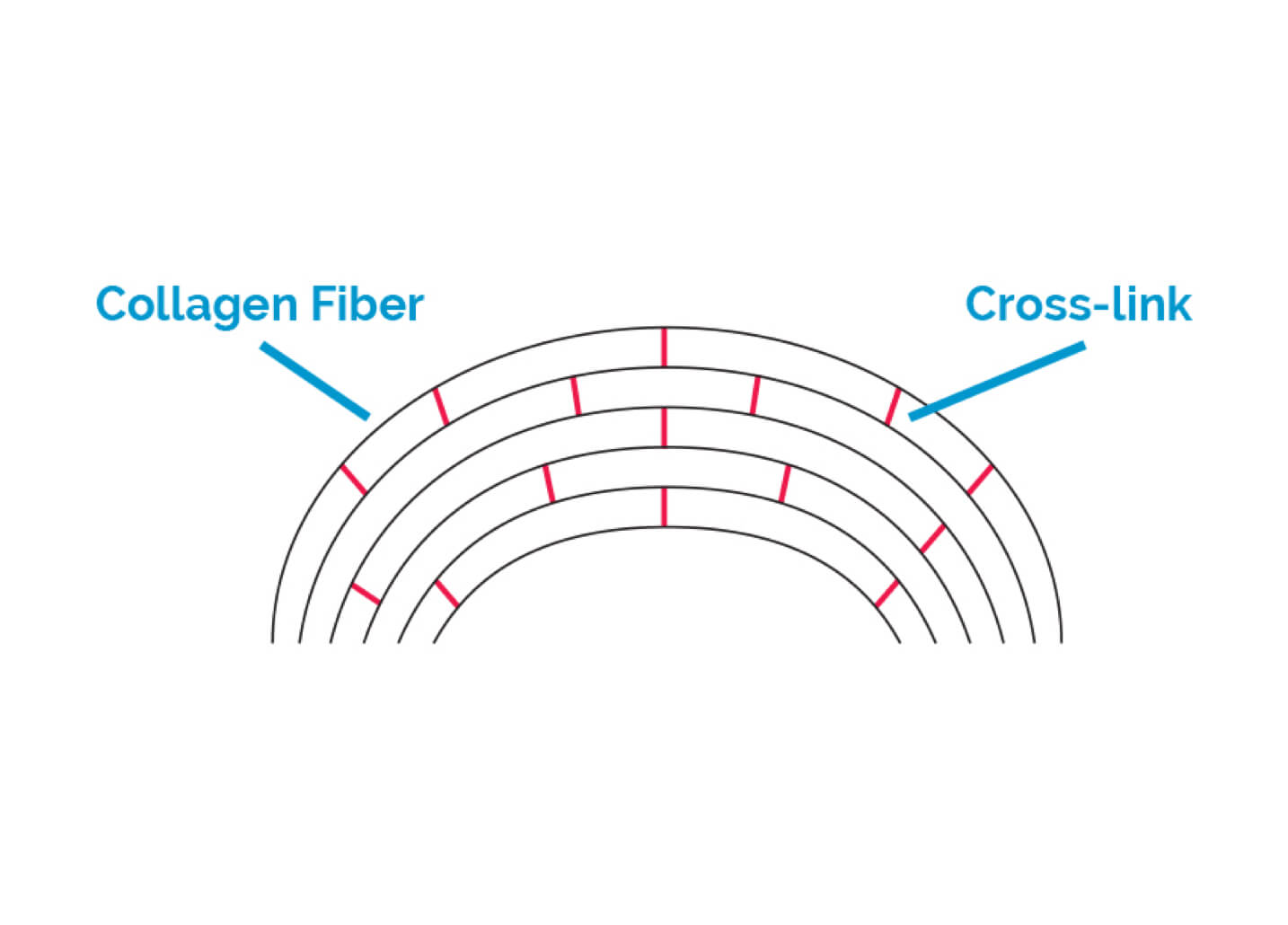
Less cross-linking leads to a weaker cornea
What is Corneal cross-linking?
Corneal cross-linking (CXL) is a treatment that strengthens the cornea and stops keratoconus from getting worse. The treatment mimics the natural cross-linking that occurs when our corneas interact with the sun’s UV rays.
Who is suitable?
CXL is recommended for patients whose keratoconus is getting worse as demonstrated by corneal curvature scans. Patients over the age of 40 generally will not require CXL as keratoconus is unlikely to progress due to the natural cross-linking that occurs with age from extended sun exposure.

UV light treatment causes new cross-links to form
How is Cross-linking performed
During the procedure, you will lie comfortably on a bed. Your eyelid will be held open with an eyelid clip. The surface cells of the cornea (the epithelium) is gently removed allowing the cornea to be soaked with Vitamin B2 (riboflavin) drops. UV light is then used to activate the riboflavin within the cornea, a process that induces cross-linking between the collagen fibres that make up the cornea, causing the cornea to become stronger.
A soft contact lens is placed over the cornea for 5-7 days at the end of the procedure.
How does cross-linking work?
CXL can be performed at MCES, in our treatment room, for those without health insurance. CXL can also be performed at the Manningham Private Hospital for those with private health insurance or when combined with laser eye surgery. The procedure is painless and takes approximately 30 minutes per eye.

More cross-linking leads to a stronger cornea
Does it work?
Studies have shown that CXL increases corneal strength by up to 300% and is successful in stopping keratoconus from getting worse in over 90% of patients. It is currently the only treatment available to stop keratoconus from worsening.
Without CXL, 20% of patients with keratoconus will eventually require a corneal transplant. However, only 3% of patients who undergo CXL will require corneal grafting.
Will it improve my vision?
The aim of CXL is to stabilise keratoconus. This treatment alone does not attempt to improve vision. After treatment, you will still need to wear your glasses or contact lenses.
If your keratoconus has progressed to a point that you have a significant refractive error or corneal scarring, other treatment options may be required to improve your vision. In some cases, this can be combined with CXL. Dr Jao will advise you of the option that is most likely to achieve the best outcome for you.
Laser vision correction

Dry Eye
Introduction
Do you suffer from dry gritty or sore eyes? You may have Meibomian gland dysfunction (MGD). Fortunately, a new technology has emerged that can stimulate Meibomian glands and reverse much of the disease. Clinical studies have shown that over 80 per cent of patients with MGD will have significant improvement in symptoms with Intense Pulse Light treatment.
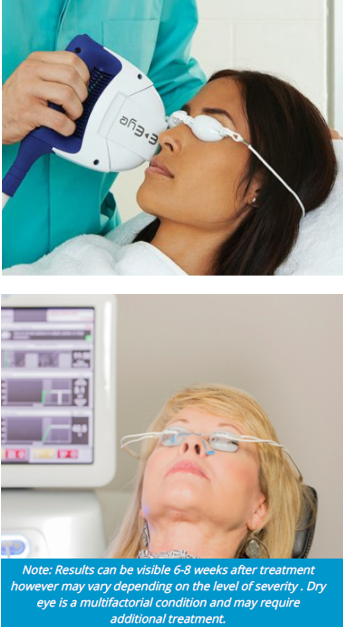
A revolutionary new treatment for sufferers of Dry Eye
What is Dry eye?
Most patients with dry gritty or sore eyes suffer from Meibomian Gland Dysfunction (MGD). If you have MGD, symptoms tent to worsen with surgical eye procedures, such as Cataract Surgery or Laser Vision Correction. Unsatisfactory vision may result after surgery if the tear film and ocular surface is not adequately managed prior.
What causes MGD?
A number of conditions can lead to changes in the structure and function of the glands, known as MGD.
Fortunately new technology has emerged that can stimulate Meibomian glands and reverse some of the disease. Clinical studies have shown that over 80% of patients with MGD who have treatment will have significant improvement in symptoms.
What is IPL?
IPL stands for Intense Pulse Light treatment, which a painless light therapy treatment. It does not require the use of injections or needles. The energy, spectrum and exposure time are precisely set to stimulate the Meibomian glands in order for them to return to their normal function. 5 ‘flashes’ along the upper cheek are given, which takes approximately 15 minutes.
What is Lipiflow?
Lipiflow treatment involves you laying comfortable on a reclined chair for 12 minutes. Anaesthetic eye drops are given. Wearing ‘ goggles’, the meibomian glands are heated and massaged to loosen the oil trapped in the glands. The softened oil glands are gently pushed up and out to express the glands and remove any blockages whilst protecting the eyes.
After treatment you may feel the eyes are very well moisturised. This can be immediate, however can take up to 8 weeks. Studies have shown that treatment effects can last up to 12 months. You can drive home after treatment.
Treatment Plan
Treatment prior to surgery can be an effective way to minimise post-surgery discomfort, therefore your Ophthalmologist may recommend you have an appointment in our specialised dry eye clinic before proceeding with surgery (fees apply). If you are not needing surgery but suffer from dry eye, we recommend a direct appointment into our dry eye clinic for a comprehensive assessment to determine what treatment option is best suited to you. If you choose to proceed with IPL, LIPIFLOW or a combination of both, it is important to continue conservative management at home.
Some patients with moderate to severe MGD/blepharitis may still benefit from routine reviews in the dry eye clinic after treatment.
Cross-Linking
Introduction
Keratoconus occurs when the cornea thins and bulges forward in a cone shape.
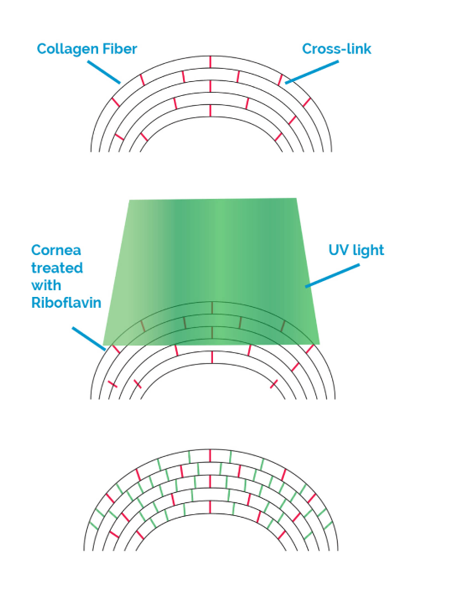
What is Corneal Cross-linking?
Corneal cross-linking (CXL) is a treatment that strengthens the cornea and stops keratoconus from getting worse. The treatment mimics the natural cross-linking that occurs when our corneas interact with the sunʼs UV rays.
Corneal cross-linking increases corneal strength by up to 300% and is successful in stopping keratoconus from getting worse in over 90% of patients. It is currently the only treatment available to stop keratoconus from worsening.
Who is Suitable?
CXL is recommended to patients whose Keratoconus is getting worse as demonstrated by corneal curvature scans.
Patients over the age of 40 generally will not require CXL as keratoconus is unlikely to progress due to the natural cross-linking that occurs with age from extended sun expose.
How is Cross-Linking performed?
During the procedure, you will lie comfortable on a bed. Your eyelid will be held open with an eyelid clip. The surface cells of the cornea (the epithelium) is gently removed allowing the cornea to be soaked with vitamin B2 (riboflavin) drops. UV light is then used to activate the riboflavin within the cornea, a process that induces cross-linking between the collagen fibres that make up the cornea, causing the cornea to become stronger. A soft contact lens is placed over the cornea for 5-7 days at the end of the procedure.
How does Cross-linking work?
CXL can be performed at MCES in our treatment room, for those without health insurance, this greatly reduced your out-of-pocket costs. CXL can be performed at the Manningham Day Procedure Centre for those with private Health insurance. The procedure is painless and takes approximately 30 minutes per eye.
Does it work?
Studies have shown that CXL increases corneal strength by up to 300% and is successful in stopping keratoconus from getting worse in over 90% of patients. It is currently the only treatment available to stop keratoconus from worsening.
Without CXL, 20% of patients with keratoconus will eventually require a corneal transplant. However, only 3% of patients who undergo CXL will require corneal grafting.
Will it improve my vision?
The aim of CXL is to stabilise Keratoconus. This treatment alone does not attempt to improve vision. After treatment, you willl still need to wear your glasses or contact lenses.
If your keratoconus has progress to a point that you have a significant refractive error, or corneal scarring, other treatment options may be required to improve your vision. In some cases, this can be combined with CXL. Dr Kathy Jao will advise you of the option that is most likely to achieve the best outcome for you.
